
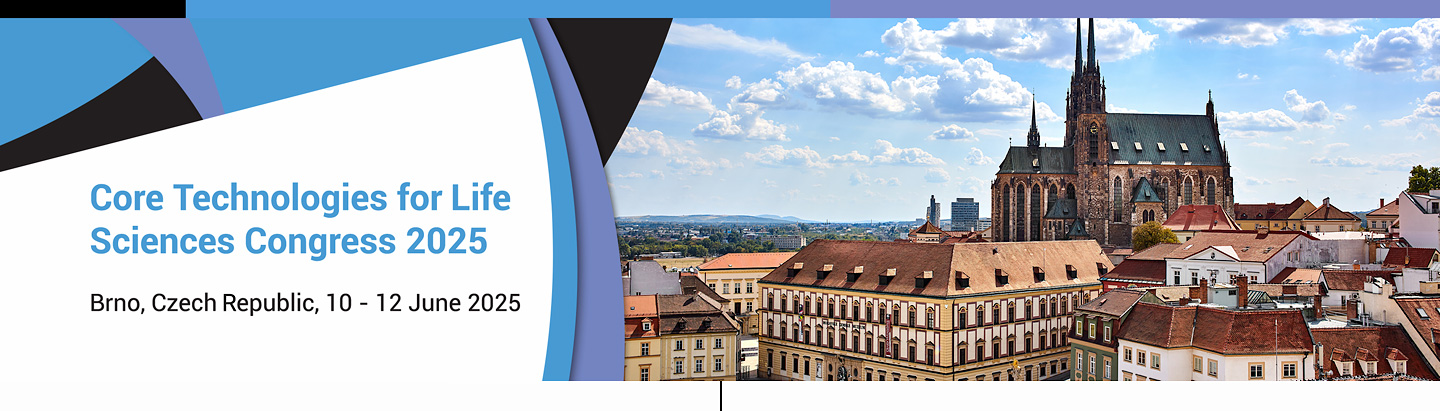
Detailed Programme
Day 1 (10 June 2025) Day 2 (11 June 2025) Day 3 (12 June 2025)
08:30
Registration open + welcome snack
HALL A+B
09:00–9:30
Welcome speech + Spencer tribute
CTLS director + CEITEC director/MU rector
HALL C+D
09:30–11:30
Congress kick-off Session: hfp consulting
HALL A+B
11:30–12:10
KEYNOTE SPEAKER TALK
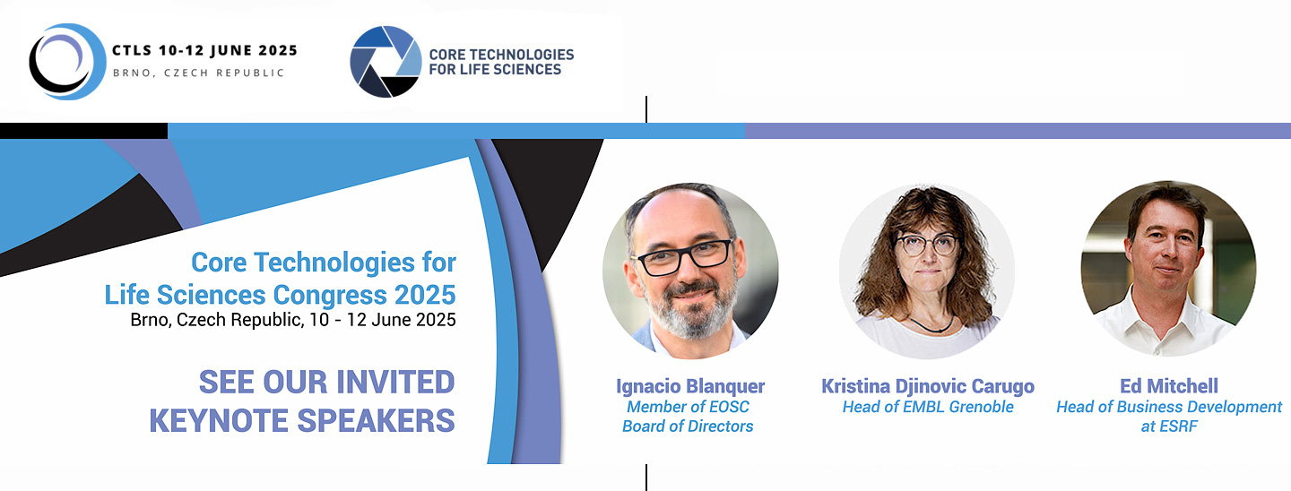
EMBL activities and the importance of core facilities for scientific institutions
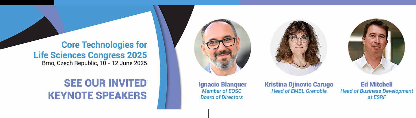
Kristina Djinovic Carugo, Head of EMBL Grenoble
12:10–12:15
Techno-byte: Beckman Coulter Life Sciences – Flow cytometry and innovation
12:15–13:15
Lunch break
12:45–13:15
Spectral Flow Cytometry Meets Modularity - CytoFLEX mosaic workshop (Beckman Coulter Company workshop)
13:15–14:15
CTLS General Assembly (open for everyone)
14:15–15:40
PLENARY SESSION 1
Co-chairs: Zuzana Hlavenková, Barry Moran
Speakers: Sonja Welsch, Sebastian Munck, Luke Brzozowski
Balancing user support and innovation in a “hands-on users” cryoEM Research Infrastructure
Sonja Welsch
Central Electron Microscopy Facility, Max Planck Institute of Biophysics, Frankfurt am Main, Germany
The operational modes of electron microscopy core facilities can vary significantly, ranging from a purely service driven mode – where expert facility staff members handle all incoming samples and operate all instruments – to a strict hands-on user mode – where all researchers are enabled by staff members to actively operate the instruments required for their projects.
A strict hands-on user mode of operation comes with pros and cons: on the one hand, it allows researchers to actively drive their own projects, control experimental conditions independently and it provides researchers with valuable hands-on expertise that can be utilized in their future career steps. it also allows facility staff members to provide support that is tailored to user needs, typically leading to a more meaningful use of instrument time. On the other hand, this mode of operation requires facility staff members to dedicate a significant portion of their capacities to training and scheduling of newly incoming users and to create and maintain training documentation. In addition, it inevitably leads to instrument use by researchers who aren’t specialists (yet) in instrument optimization and troubleshooting.
We will discuss how the Central Electron Microscopy facility at Max Planck Institute of Biophysics strives to balance the pros and cons of such a hands-on user mode of operation, to optimize both quality and quantity of users’ results and how to balance innovation and continuity of electron microscopy workflows by close collaboration with soft- and hardware suppliers as well as facility users.
From development to service, opportunities for imaging facilities
Sebastian Munck
1VIB Bio Imaging Core, VIB, Leuven, Belgium; 2VIB Center for Brain & Disease Research, VIB, Leuven, Belgium; 3Department of Neurosciences, KU Leuven, Leuven, Belgium
Core facilities are vital for providing services to departments and stakeholders, but without innovation, they risk stagnation and failure to advance science. Core facilities are often based at discovery-based life science institutes, where development is a distinct activity and where a development project within a core facility is a temporary endeavor to produce a unique product, service, or result, turning a challenge into a solution. These projects vary in complexity, ranging from complete new imaging methods to protocol optimizations and technology adaptation.
For imaging core facilities, different opportunities for innovation do exist. Here, we will review different possibilities and focus on investigating comparative approaches for multiscale three-dimensional imaging. We showcase this approach with an example of murine lung vasculature from macro- to micro-structural level, comparing different imaging techniques and the results they yield. We believe that this is a crucial exercise for a core facility to be able to advise users on the best solution for their scientific projects. Consequently, we will highlight some aspects of our efforts to establish a data journey for our users and discuss our efforts to interact with other infrastructures, such as the Flemish supercomputing center, for that purpose.
Overall, we believe that establishing workflows and committing staff time to innovative/development activities that benefit the majority of users is essential for creating scalable development projects for long-term service implementation. A structured approach with a road map with go-and-no-go decisions and milestones can make this process transparent and tangible for the facility’s stakeholders.
Facilitating Innovation at an Academic Hospital
Luke Brzozowski
Executive Director, Translational Research and Core Faculties; University Health Network, Toronto, Canada
University Health Network (UHN) is affiliated with the University of Toronto and consists of Toronto General and Toronto Western Hospitals, the Princess Margaret Cancer Centre, Toronto Rehabilitation Institute, and The Michener Institute of Education at UHN. The scope of research and complexity of cases at University Health Network has made it a national and international source for discovery, education and patient care. It has the largest hospital-based research program in Canada, with major research in arthritis, cardiology, transplantation, neurosciences, oncology, surgical innovation, infectious diseases, genomic medicine and rehabilitation medicine.
I will describe several mechanisms that have been put in place at UHN to facilitate translational innovation. First I will talk about UHN Research and Innovation Cores, a uniform approach towards enabling UHN researchers’ and external users access to over 20 state-of-the-art facilities and service groups across areas as diverse as genomics, flow cytometry, medical imaging, engineering, or biobanking services. Then I will dive deeper into a specific core, the Techna Technology Development Team. Techna consists of 50 technical project managers, engineers, and software developers who lead, manage, and carry out health technology productization programs and projects in a hospital environment. Lastly, I will conclude with a description of a particular translational program – the Theranostics Program and UHN. I will use this example to illustrate how we approach organizing innovation by using our core facilities, navigating policy makers, philanthropy, clinical teams, and private sector to bring this new fascinating diagnosis and treatment platform from bench to bedside.
15:40–15:45
Techno-byte: agendo.science – Give me five! Or just one
15:45–16:15
Coffee break
16:15–17:40
PLENARY SESSION 2
Co-chairs: Julia Fernandez Rodriguez, Pavel Tomančák
Speakers: Fabia Gozzo, Petr Strnad, Vendula Hrabalová, Vladimír Zelník
Bridging the Innovation Gap: Industry-Focused Synchrotron XRPD Solutions for Pharmaceutical and Chemical Challenges
Fabia Gozzo
Excelsus Structural Solutions (Swiss) AG, PARK INNOVAARE, Parkstrasse 1, 5234 Villigen, Switzerland
Synchrotron-based X-Ray Powder Diffraction (XRPD) is redefining structural analysis, providing the pharmaceutical and chemical industries with unmatched precision and capability for material characterization. This presentation will highlight how Excelsus Structural Solutions enables industrial customers to leverage advanced structural characterization by integrating synchrotron methodologies—without requiring them to navigate the complexities of direct facility access. Our innovations in XRPD methodologies achieve detection and quantification thresholds as low as 0.01%wt for crystalline phases in organic pharmaceutical compounds, making these capabilities readily available to industry partners, who can benefit from precise structural analysis without engaging directly with synchrotron instrumentation and technology. These advanced methodologies also allow for amorphous quantification without the need to spike the compound under investigation. By introducing refined calibration techniques—unaffected by inhomogeneous phase distribution within samples—our XRPD methodologies not only meet rigorous industrial standards for quantification but also exceed industry benchmarks.
The talk will showcase successful technology transfer, bridging gaps in knowledge exchange between complex research infrastructures and industry, and emphasizing real-world case studies. These examples demonstrate the impact of custom-tailored synchrotron solutions in overcoming industrial analytical challenges, enhancing both sensitivity and efficiency in structural characterization. The session will underscore how strategic research-industry partnerships not only advance technology transfer but also address critical needs in drug development and chemical analysis, accelerating innovation with practical, scalable solutions to develop higher-quality products.
Viventis Microscopy: Developing advanced light sheet microscope systems for live imaging of large multicellular systems
Petr Strnad
Viventis Microscopy Sàrl, Lausanne, Switzerland
Light sheet fluorescence microscopy (LSFM) is a preferred method for imaging living samples due to its high imaging speed and low phototoxicity. Organoids, which are 3D multicellular model systems that mimic the structure and function of organs in vitro, present a challenge for imaging as they develop over several days and grow to large sizes.
Viventis Microscopy was established to develop and commercialize light sheet microscope systems specifically designed for live imaging of such systems. To address the challenges of imaging large samples, SiMView, MultiView, and Panoramatic LSFM have been proposed. These systems use an identical concept: the sample is viewed from two opposing directions using two detection objectives and illuminated from two opposing directions using two illumination objectives. This setup increases the penetration depth by a factor of two for optimally sized samples. However, the sample must be held in a constrained space between the four objectives, making multi-position and organoid imaging difficult.
In collaboration with Prisca Liberali's group at FMI in Basel, Viventis Microscopy has developed an open-top dual-view and dual-illumination light-sheet microscope designed for live imaging of large specimens. This novel design overcomes the limitations of traditional setups. It is using two opposing detection objectives to image sample from two directions and two illumination objectives which are opposing but slightly rotated to point upwards. This creates an unobstructed linear space just above the two illumination objectives for a custom designed sample holders containing a linear array of wells and samples for multi-position imaging.
Low-Voltage Transmission Electron Microscopy for Biopolymer Imaging
Vendula Hrabalová1, Jaromír Bačovský1, Kateřina Mrázová2, Eva Slaninová3
1Delong Instruments a.s., Brno, Czech Republic; 2Institute of Scientific Instruments of the CAS, Brno, Czech Republic; 3Faculty of Chemistry, Brno University of Technology, Brno, Czech Republic
Low-voltage transmission electron microscopes (LVEMs) are advanced imaging techniques with many applications in materials and life sciences. LVEMs have also found valuable applications in biopolymer imaging because they offer high-contrast visualization without the necessity of using toxic staining agents. High-voltage TEMs struggle with low-contrast imaging of biopolymers due to their composition of light elements. LVEMs operating at 5–25 kV increase electron scattering, naturally increasing contrast and preserving sample integrity.
This study highlights the advantages of LVEMs, particularly in imaging polyhydroxyalkanoates (PHAs), group of biocompatible and biodegradable polyesters produced by some microorganisms in the form of intracellular granules. PHAs have properties similar to petroleum-based plastics, making them suitable substitutes for packaging materials. PHAs can also be used in agriculture (e.g., mulch films), medical applications (e.g., sutures and tissue engineering), and 3D printing. High voltage TEM methods often require staining with uranyl acetate or osmium tetroxide, which may cover fine structural details of samples and pose health risks. LVEMs eliminate this requirement and allow for the direct visualization of the samples.
The All-in-One Electron Microscope LVEM 25E integrates TEM, SEM, STEM, ED, and EDS modalities within a single system, providing a comprehensive multimodal approach to biopolymer research. This combination enhances imaging versatility. STEM mode further improves image sharpness by minimizing the effect of chromatic aberration, which provides better contrast for thicker samples. In addition, the ability to work with standard sample thicknesses rather than requiring ultrathin sections makes this approach a practical tool for routine imaging of biological and material samples.
Biotechnology Laboratories of the Biomedical Research Centre, Slovak Academy of Sciences - Activities and Cooperation Opportunities
Vladimír Zelník, Marta Miklošová, Juraj Kopáček
Biotechnology Laboratories, Biomedical Research Centre, Slovak Academy of Sciences, remote site in Šarišské Michaľany, Slovak Republic
The main objective for the founding of Biotechnology Laboratories (BTL) of the Biomedical Research Centre, Slovak Academy of Sciences has been to provide an effective means for the transfer of research results on biopharmaceuticals into practice utilizing novel approaches and modern infrastructure.
Slovakia is among EU countries lagging in its capability to introduce results of medical and pharmaceutical research into general practice and as consequence also in the contribution of pharmaceutical production to the country’s economy. The creation of BTL opened possibilities for applied research on the introduction of novel approaches to the development, production, purification and characterization of biologically active macromolecules and multicomponent substances for therapeutic, preventive and diagnostic purposes and their effective quality control.
The BTL consist of Research Laboratories for the development and preparation of biopharmaceuticals and Analytical Laboratories.
Research Laboratories provide space for the development of therapeutic biopharmaceuticals produced in eukaryotic cell systems, novel viral vaccines with a focus on their better efficacy and safety and therapeutic and diagnostic substances of bacterial origin.
Analytical Laboratories provide opportunities for the development, validation and accomplishment of analytical methods for testing of efficacy of newly developed biologically active substances, definition of their composition, identity, purity and safety. BTL Analytical Laboratories carry GMP certificate for testing laboratories in pharmacy.
We will present actual examples how the BTL carry out its R&D activities on basis of research projects supported by grant schemes and collaboration with Slovak academic institutions, as well on basis of contractual research and analyses for commercial pharmaceutical enterprises.
17:40–17:45
Techno-byte: Idea Elan - Manage all your cores in a few clicks with Infinity X
17:45–18:15
Infinity X: Seeing is believing! (Idea Elan Company workshop)
17:45–19:30
Entertaining programme in Brno
19:30–21:00
Welcome drink and Poster Session (Hall C)