
Preliminary Detailed Programme
Day 1 (10 June 2025) Day 2 (11 June 2025) Day 3 (12 June 2025)
08:30
Registration open
HALL A+B
09:00–09:45
KEYNOTE SPEAKER TALK
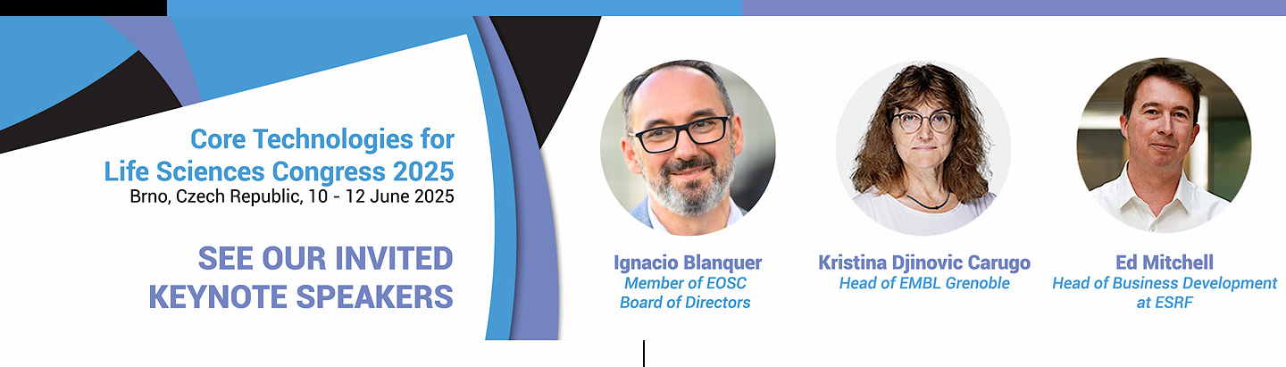
Ed Mitchell, Head of Business Development at ESRF
The European Synchrotron Radiation Facility: a knowledge hub for industry
Synchrotron light sources the world over are driving forwards innovation thanks to their unique capabilities provided by intense X-ray beams. Stakeholders are demanding increased economic return and part of this is a stronger engagement with industry. The European Synchrotron Radiation Facility (ESRF) is no exception to this and is integral to advancing life sciences by actively engaging with both academic and industrial partners. Offering full access to all beamlines and services for publishable research and commercial innovation, ESRF provides a unique platform for cutting-edge research. This presentation will delve into how ESRF’s advanced synchrotron technologies, particularly the Extremely Brilliant Source (EBS), are harnessed in collaboration with industry to drive innovation in life sciences. Key projects will be highlighted, illustrating the application of these technologies in areas such as drug discovery, structural biology and biotechnology, as well as from areas outside of life sciences and where new services have been created and tailored for industry. Additionally, the presentation will address the barriers and challenges faced in these collaborations, looking at the complexities of integrating synchrotron science into industrial research and development.
09:45–11:00
OPEN MIC SESSION
Co-Chairs: Josef Houser, Thomas Trüb
11:00–11:30
Coffee break
11:30–13:00
PLENARY SESSION 8
Co-Chairs: Aleš Benda, Josh Rappoport
Speakers: Johanna Bischof, Fernando Pelaez, Krzysztof Skowronek
Euro-BioImaging – facilitating multimodal imaging across Europe
Johanna Bischof
Euro-BioImaging ERIC
Euro-BioImaging – as a European Research Infrastructure – offers all scientists open access to a large portfolio of imaging instruments, expertise, training opportunities, and image data services. The technologies offered by Euro-BioImaging can be accessed at Euro-BioImaging Nodes, which comprise more than 200 internationally renowned imaging facilities distributed across Europe. They cover the whole spectrum of biological and biomedical imaging, from the molecular to the human scale. In addition to access to cutting-edge instruments, the Euro-BioImaging Nodes provide expertise, guidance and training on all aspects of the imaging experiment – from experimental design and sample preparation to image analysis services, including on external data.
All scientists, regardless of affiliation, area of expertise, or field of activity, can benefit from Euro-BioImaging’s pan-European open access services and funding for user access is available.
With its broad portfolio of imaging technologies and consortium of expert facilities, Euro-BioImaging actively supports researchers in applying multimodal and correlative imaging to their research question.
For the technical experts at imaging core facilities, Euro-BioImaging provides a cross-European network and platform for exchange of experience, as well as new training opportunities. This network of technical experts facilitates cross-facility collaborations, imaging workflows, and technical innovations.
How to make easier to include several core facilities in research projects?
Fernando Peláez
Biotechnology Programme, Spanish National Cancer Research Centre (CNIO), Madrid, Spain
Cutting-edge biomedical research relies heavily in the use of increasingly complex technologies. Multidisciplinary approaches to a given biological problem have become the norm and are usually demanded by top journals to warrant publication of research results. In this context, CFs play a critical role in research institutions to ensure high-quality access to those technologies. However, we often see that the outstanding potential of CFs is not fully realized and there are projects that do not take all the advantage from such diversity of approaches and technologies, which often involves several CFs working in a coordinated manner. Why is this the case?
Experimental design is usually done by researchers with a limited knowledge of technologies, or with a strong bias towards any particular discipline. They may or may not be aware of the potential advantage or applications of some technologies to address their questions of interest. CF heads can play an essential role to fill that gap of knowledge. It is of paramount importance to generate a culture in the organizations by which the input and advice of CF heads can be gathered early enough in the life cycle of projects to ensure optimal planning, as well as coordination among the CFs. Likewise, it is important to have a flexible organization that facilitate the cooperation of different CFs working on the same project without impediments.
Some examples of successful projects ran at CNIO which took advantage of multiple technologies involving different CFs will be described.
Cross-Platform Collaboration to Develop New Models for Rare Diseases in IN-MOL-CELL infrastructure
Krzysztof Skowronek, Olga Gewartowska, Andrzej Dziembowski
Molecules and Cells Research Infrastructure (IN-MOL-CELL), International Institute of Molecular and Cell Biology in Warsaw (IIMCB), Warsaw, POLAND
We present the Institute of Molecular and Cell Biology (IIMCB)’s plans to create the coordinated services of multiple Core Facilities within the IN-MOL-CELL infrastructure to support research efforts in the RACE Prime project. This initiative aims to develop novel models for rare diseases under investigation at our Institute.
Each project begins with a research group proposing one or more target genes that were identified by the collaborating team at the paediatric hospital as linked to a specific rare disorder. The integrated pipeline carried out by IN-MOL-CELL facilities starts with introduction of precise genetic modifications into a range of model organisms, including human cells, mice, zebrafish, and C. elegans. Human cells are further utilized to generate relevant organoid models.
In vivo models (mice, zebrafish, and C. elegans) are characterized for behavioral and physiological alterations (in specialized CFs), while tissue samples are screened using high-content screening platforms, along with more specialized microscopy techniques when required. To obtain a comprehensive understanding, selected types of cells from will be isolated and undergo in-depth analysis via RNA sequencing, proteomics, and metabolomics. In select cases, protein products of genes of interest will be purified and characterized using structural biology and biophysical methods.
The ultimate goal of this coordinated effort across all 10 facilities in IN-MOL-CELL is to deliver a comprehensive set of highly relevant disease models with multi-layered characterization. To support this ambitious, high-risk initiative, our Institute has secured its place on the national research infrastructure roadmap, alongside various funding sources to finance IN-MOL-CELL operations.
13:00–14:00
Lunch break
14:00–15:15
PLENARY SESSION 9
Co-Chairs: Monica Morales, Vivian Lu Tan
Speakers: Simon Devos, Milena Hasan, Sebastian Munck, Andreas Sommer
QSample Project
Simon Devos1, Eduard Sabido2, Julia Ponomarenko3
1VIB Proteomics Core, VIB, Ghent, Belgium; 2Proteomics Unit, CRG, Barcelona, Spain; 3Bioinformatics Core, CRG, Barcelona, Spain
To assure the reliable and reproducible generation of mass spectrometry-generated proteomics data, standardized quality control procedures are essential. Ideally, these procedures are implemented throughout different proteomics core facilities and research groups to enable inter-laboratory harmonization in defining optimal performance conditions of both LC and MS infrastructure. In this session we will elaborate on some QC initiatives across core facilities to enhance data comparability and reliability.
After the success of the QCloud system1,2,3 to monitor the performance of mass spectrometers within the Core for Life proteomics laboratories, the proteomics and bioinformatics units from the Centre for Genomic Regulation (CRG) and the Flemish Institute for Biotechnology (VIB) joined forces to develop an automated quality control system for proteomics experiments and actual user samples. Based on a prototype system called QSample developed in the CRG Proteomics Unit, the goal is to develop a system that automatically monitors the key quality indicators of each sample processed in a proteomics laboratory, which can be adopted to the different Proteomics Units of the Core for Life alliance, increasing overall quality and efficiency of LC-MS/MS analysis of real user samples.
1Chiva C et al (2018). QCloud: A cloud-based quality control system for mass spectrometry-based proteomics laboratories. PLoS One.
2Olivella R et al (2021). QCloud2: An Improved Cloud-based Quality-Control System for Mass-Spectrometry-based Proteomics Laboratories. J Proteome Res.
3Chiva C et al (2025). A Multiyear Longitudinal Harmonization Study of Quality Controls in Mass Spectrometry Proteomics Core Facilities. J Proteome Res.
Inter-institutional benchmarking of the Visium 10XGenomics pipeline
Milena Hasan, on behalf of the C4L Single Cell Task Force
Single Cell Biomarkers UTechS, C2RT, Institut Pasteur, Paris, France
Spatial transcriptomics is the latest layer of single-cell multi-omics. It allows transcriptomic profiles to be linked to their tissue localization, opening up the possibility of studying the relationship between gene expression signatures in individual cells and the cellular microenvironment.
There are two types of technologies for spatial transcriptomics analysis. NGS-based pipelines, such as Visium (10X Genomics) and Stereo-Seq (BGI), rely on sequencing as the readout, allowing the study of the entire cellular transcriptome through polyA capture of RNA. In contrast, microscopy-based instruments such as MERSCOPE (Vizgen) and Xenium (10X Genomics) are used to quantify fluorescently labelled target transcripts.
The first commercialized pipeline was Visium, which remains the most widely used technology for spatially resolved transcriptomics. Despite its application in basic and biomedical research projects since 2019, there is a lack of interlaboratory benchmarking of the Visium pipeline. In addition, the susceptibility of different steps of the pipeline to technical error and bias introduction has not been fully addressed.
In this work, we used a mouse model of Alzheimer's disease to perform an inter-laboratory benchmarking of the Visium pipeline between core facilities of six C4L institutes. After centralized tissue preparation, downstream sample processing, library preparation, qualification, and sequencing were performed by the partner laboratories. Data analysis was performed by each partner and integrated into a centralized analysis.
The establishment of the project and the results obtained will be discussed.
Best practices from the community for the community – prototyping within the Core for Life Microscopy Workgroup
Sebastian Munck
BioImaging Core Leuven, VIB, Belgium; the C4L Microscopy Workgroup; and the C4L consortium
Scientific progress in life sciences relies on expanding technologies, expertise, multidisciplinary approaches, and collaborations rather than a single-laboratory environment. Consequently, the scientific core facility model has been developed to address this issue, with distinct central facilities serving a broad base of users and focusing on specific techniques. However, collaboration goes beyond the single institute and consequently requires best practices and standard procedures based on a dialog with peers. Imaging and microscopy core facilities are abundant due to their widespread demand.
To address the challenges of maintaining momentum and high-quality daily operations in a high-end scientific imaging core facility, the Core for Life microscopy workgroup has created a resource to address these tasks. This resource includes ideas for maintaining momentum, managing equipment and users, handling repairs and service contracts, planning for equipment upgrades, renewals, or decommissioning, and continuously upskilling while balancing innovation and consolidation (Reynaud et al., 2024).
Another more recent resource explores the effects of working in highly competitive environments and dealing with stressful situations. The work includes questionnaires on responding to stressful situations, conflict management, and establishing a peer-led mutual-aid group focused on mental health. This resource may be valuable for the broader CTLS public and all 'walk-in' facilities, such as flow and related ones.
The crosstalk and cross-fertilization between communities like C4L and CTLS will be discussed using these examples.
Can we fix them all? A benchmarking study on the suitability of cell fixation across multiple single-cell platforms
Thomas Grentzinger1, Philip Wolff1, Hubert Rehrauer2, Catharine Aquino2, Andreas Sommer1
1NGS Core Facility, Vienna Biocenter Core Facilities (VCBF), Vienna, Austria; 2 Genomics Core, Functional Genomics Center Zurich (FGCZ), Zürich, Switzerland
Single-cell RNA sequencing (scRNA-seq) is a powerful approach that allows the examination of the transcriptome of individual cells within heterogeneous biological samples. However, the traditional gold standard workflows were designed for freshly dissociated single cell suspensions that i) entail a flawless organization and execution of the experiment, ii) are subject to cellular stress bias due to the intrinsically transient nature of the transcriptome and iii) in some case strongly limit or even prohibit specific experimental designs due to trivial logistic considerations.
To address these limitations, the present project seeks to evaluate the efficacy of commercially available and reliable cell fixation protocols and storage methods across various scRNA-seq platforms accessible within our facilities. Reliable cell fixation will allow us to disconnect the initial sample generation from all downstream processing steps.
Validating a cell fixation and storage procedure compatible with the currently applied scRNA-seq platforms has manifold benefits: improving access to scRNA-seq and the quality of the data generated as well as optimizing and securing the workflows our scientific community relies on.
15:15–15:30
Closing remarks
CTLS president + representative of local organizer